
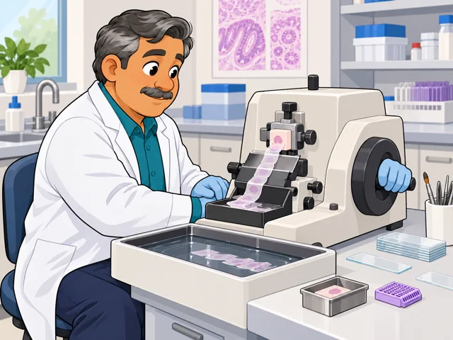
Embed tissue, then slice it
You set tissue in a wax block first, then cut very thin slices for slides on a microtome.
Career profile
Histotechnologists make fine tissue detail show clearly and find out why a slide went wrong.
Want precise lab work that puts biology in your hands? You prepare tissue for pathologists, troubleshoot machines and judge how well lab methods work.
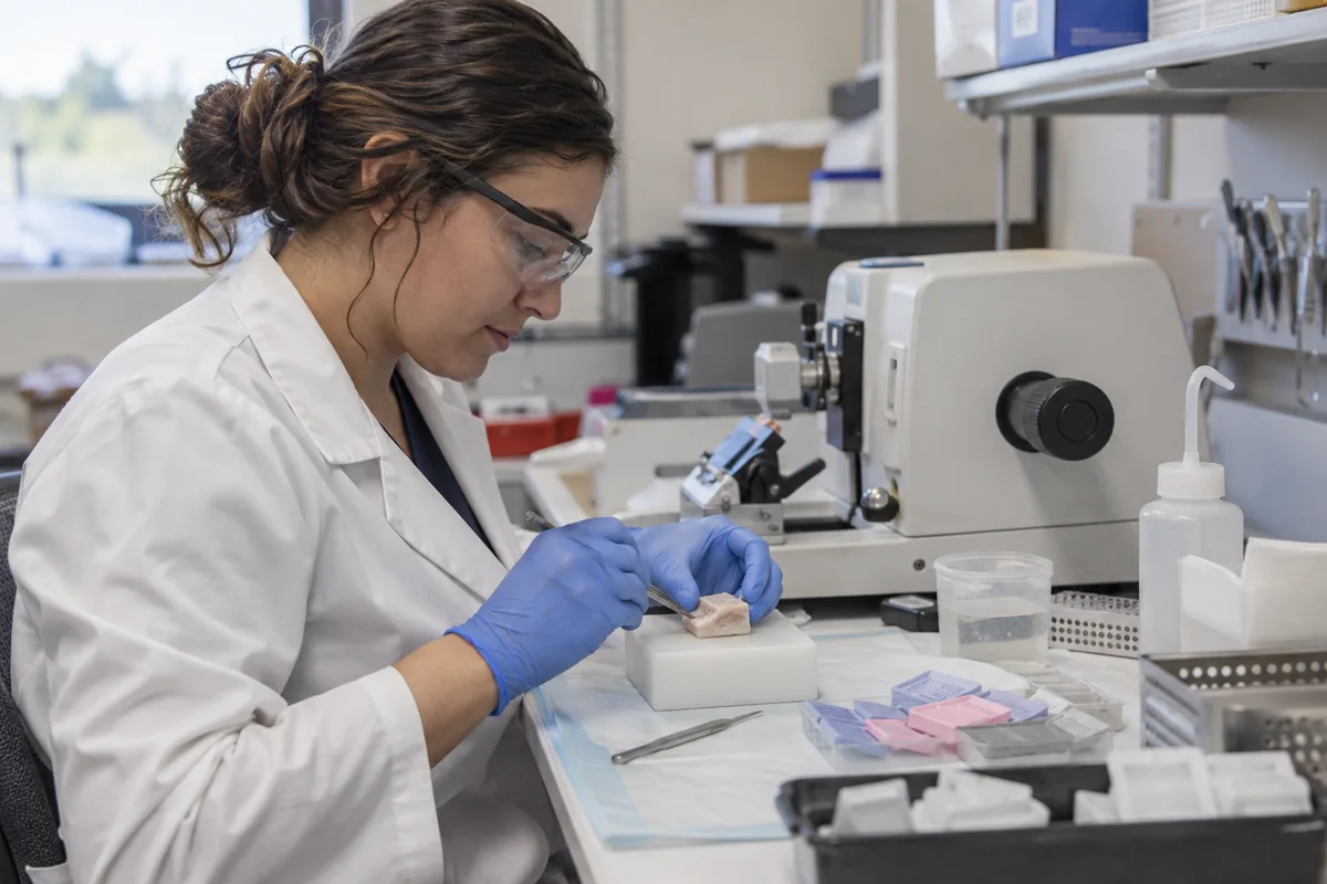
You set tissue in a wax block first, then cut very thin slices for slides on a microtome.
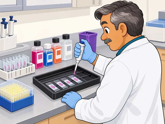
Apply stains so structures show up, and in specialist immunohistochemistry work, mark specific proteins with antibodies.
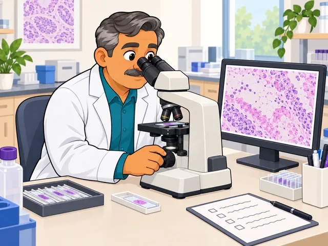
If staining looks off under the microscope, check slide prep, stain controls and equipment before anyone accepts results.
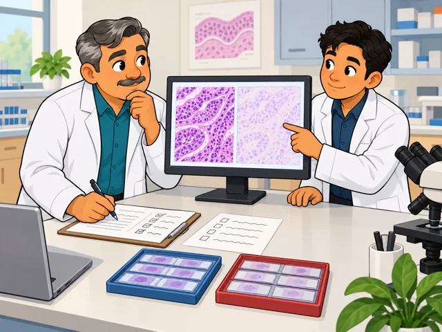
In some positions, you help test a new staining method, measure it against lab needs and note problems.
United States · 2026
A national median is not reported for this title. Broader clinical laboratory pay appears below.
National growth and openings are not reported separately for histotechnologists.
Quick orientation
The certification routes below pair a degree with accredited training or qualifying laboratory experience.
A national median is not reported for this title. Broader clinical laboratory pay appears below.
National growth and openings are not reported separately for histotechnologists.
Automated instruments can run staining steps. In some labs, scanners turn glass slides into digital images for viewing and analysis.
You still maintain instruments, investigate failed runs and check whether a method produces usable results.
How much troubleshooting can become automated remains uncertain, especially when unfamiliar tissue or a new method behaves differently.
Pay in context
Histotechnologist-specific national pay is not reported. The broader clinical laboratory workforce offers context, but its figures are not a salary estimate for this specialty.
Clinical laboratory technologists and technicians had a median of $62,930 a year, or $30.26 an hour, in May 2025. The lowest 10% earned below $38,910 annually; the highest 10% earned above $100,990. These are wage percentiles, not starting and senior salaries.
For clinical laboratory technologists and technicians overall, employment is projected to grow 3% from 2025 to 2035, with about 20,800 openings annually. Many replace people who leave the occupation. Histotechnologists' share is not reported.
An employer may weigh your experience, skills and education when setting pay. Check whether a quoted range excludes overtime, shift differentials and benefits.
Two routes lead toward the American Society for Clinical Pathology (ASCP) Scientist in Histotechnology certification, HTL(ASCP). After meeting eligibility requirements, you must pass the exam.
After entry
This starts where the entry routes leave off: early work, growing capability, and advancement or specialization. This path also has a credential or licensing boundary; see the requirements and limits.
Learn your lab's specimen records and workflow. Build consistency in embedding, cutting and staining before taking on unfamiliar preparations.
Develop skill in specialized staining and tracing technical failures. Learn how to assess a method's performance, not just complete its steps.
A senior histotechnologist can help validate methods, tackle difficult troubleshooting and coach colleagues. This adds responsibility for how the laboratory's work holds up.
You may work within a specialty team handling surgical specimens, frozen sections or specialized stains. High specimen volume shapes the pace; some positions include off-shifts, weekends or on-call work.
You prepare tissue for investigators' experiments. A project may need sections cut to particular anatomy or different preparation methods; some cores also stain and scan slides. Discussing experimental goals helps determine the work.
Try it
Choose an openly available teaching image of a stained tissue slide. Make a blurred or low-contrast copy, then compare them: which details became harder to see? Write two questions you would ask about slide preparation. Notice whether checking image quality makes you curious about the process behind it.
Stay safe: Use teaching images only; this is a quality-observation exercise, not diagnosis or specimen handling.